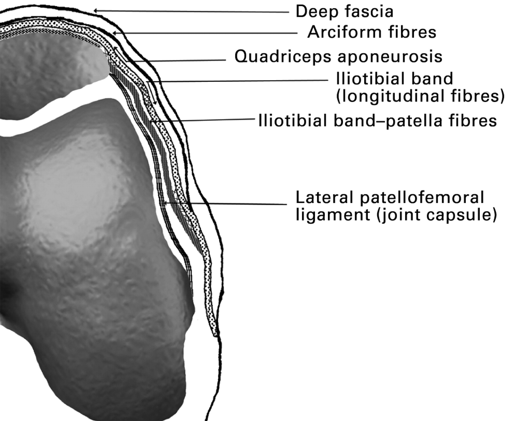
Anatomy of the Medial Retinaculum

The medial retinaculum is a fibrous band that forms the medial border of the carpal tunnel. It originates from the pisiform bone and triquetrum and inserts into the hook of the hamate and the base of the fifth metacarpal. The medial retinaculum serves to hold the tendons of the flexor carpi radialis, palmaris longus, and flexor carpi ulnaris in place as they pass through the carpal tunnel.
The medial retinaculum, a connective tissue structure in the wrist, plays a crucial role in stabilizing the carpal bones. It’s like a master conductor, ensuring harmony among these small bones. And just as Al Horford , the legendary basketball player, seamlessly orchestrates his team’s offense, the medial retinaculum deftly coordinates the wrist’s movements, allowing us to perform countless daily tasks with ease.
Structure, Medial retinaculum
The medial retinaculum is a thick, fibrous band that is approximately 2 cm wide and 1 cm thick. It is attached to the pisiform bone and triquetrum proximally and to the hook of the hamate and the base of the fifth metacarpal distally. The medial retinaculum is continuous with the transverse carpal ligament, which forms the roof of the carpal tunnel.
The medial retinaculum, a ligament that stabilizes the wrist, can be strained by repetitive movements, like those performed by basketball legend James Worthy. This ligament helps prevent excessive bending and twisting of the wrist, ensuring its smooth and pain-free function.
Function
The medial retinaculum serves to hold the tendons of the flexor carpi radialis, palmaris longus, and flexor carpi ulnaris in place as they pass through the carpal tunnel. This prevents the tendons from bowing or buckling, which could lead to pain and inflammation.
Clinical Significance
The medial retinaculum can become inflamed or thickened, which can lead to carpal tunnel syndrome. Carpal tunnel syndrome is a condition that causes pain, numbness, and tingling in the hand and forearm. It is caused by compression of the median nerve, which passes through the carpal tunnel. Treatment for carpal tunnel syndrome typically involves splinting, corticosteroid injections, or surgery.
Medial Retinaculum Injuries

The medial retinaculum is a strong band of connective tissue that runs along the medial side of the wrist. It helps to stabilize the wrist joint and prevents the tendons from bowing out. Medial retinaculum injuries can occur due to overuse, trauma, or a combination of both.
Symptoms of medial retinaculum injuries can include pain, swelling, and tenderness along the medial side of the wrist. The pain may be worse with certain activities, such as gripping or twisting the wrist. In severe cases, the medial retinaculum may rupture, which can lead to instability of the wrist joint.
Types of Medial Retinaculum Injuries
There are several different types of medial retinaculum injuries, including:
- Sprains are injuries to the ligaments that connect the medial retinaculum to the bones of the wrist. Sprains can range from mild to severe, depending on the severity of the injury.
- Strains are injuries to the tendons that pass through the medial retinaculum. Strains can also range from mild to severe, depending on the severity of the injury.
- Tears are complete ruptures of the medial retinaculum. Tears are usually caused by a sudden, forceful injury to the wrist.
Treatment and Recovery
The treatment for medial retinaculum injuries depends on the severity of the injury. Mild injuries can often be treated with rest, ice, and compression. More severe injuries may require surgery to repair the damaged tissue.
The recovery time for medial retinaculum injuries varies depending on the severity of the injury. Mild injuries may heal within a few weeks, while more severe injuries may take several months to heal.
Medial Retinaculum Release Surgery

Medial retinaculum release surgery is a procedure performed to alleviate pain and discomfort caused by the compression of the median nerve within the carpal tunnel. This surgery involves cutting the medial retinaculum, a ligament that forms the roof of the carpal tunnel, to create more space for the median nerve and its surrounding tendons.
Indications for Medial Retinaculum Release Surgery
- Persistent pain and numbness in the thumb, index, middle, and ring fingers
- Weakness in the thumb muscles, making it difficult to perform tasks such as gripping or pinching
- A positive Tinel’s sign, which involves tapping over the median nerve at the wrist and eliciting a tingling sensation in the fingers
- A positive Phalen’s test, which involves flexing the wrists for a minute and experiencing numbness or tingling in the fingers
- Failure of conservative treatments, such as splinting, corticosteroid injections, or activity modification
Surgical Procedure
Medial retinaculum release surgery is typically performed under local anesthesia. The surgeon makes a small incision in the palm of the hand at the wrist crease. The medial retinaculum is then identified and carefully divided, creating more space for the median nerve and tendons. The incision is closed with sutures, and a bandage is applied.
Risks and Benefits
As with any surgical procedure, medial retinaculum release surgery carries certain risks and benefits:
Risks:
- Infection
- Bleeding
- Nerve damage
- Scarring
- Failure to relieve symptoms
Benefits:
- Relief from pain and numbness
- Improved hand function
- Increased range of motion in the wrist and fingers
- Prevention of further nerve damage
Recovery and Rehabilitation
After medial retinaculum release surgery, the hand is typically immobilized in a splint or cast for a few weeks. Physical therapy is often recommended to help restore range of motion, strength, and function to the hand. Most patients experience significant improvement in their symptoms within a few months after surgery.
The medial retinaculum, a connective tissue in the wrist, plays a crucial role in stabilizing the carpal bones. Its significance extends beyond the realm of anatomy, as its name resonates with the poignant question: how did Jerry West die ?
While the circumstances surrounding the legendary basketball player’s passing remain a mystery, the medial retinaculum serves as a reminder of the fragility of life and the enduring impact of our actions on the tapestry of history.
The medial retinaculum, a crucial ligament in the wrist, plays a vital role in stabilizing the carpal bones. Its intricate structure resembles a delicate lacework, connecting to various bones and muscles. This intricate ligament echoes the complex ownership structure of the Mavericks, where a group of investors, including mavs owner Mark Cuban, share the reins of the team.
Like the medial retinaculum, their collective efforts guide the Mavericks towards success, ensuring the team’s resilience and agility on the court.
The medial retinaculum, a band of connective tissue that stabilizes the flexor tendons in the wrist, shares a remarkable connection with the philanthropic legacy of Miriam Adelson. Just as the retinaculum provides a supportive framework for the tendons, Adelson’s generosity has nurtured countless lives through her support of medical research and education.
And just as the retinaculum’s delicate balance ensures proper hand function, Adelson’s philanthropic vision has fostered a vibrant ecosystem of innovation and compassion.